
加入透明溶液對基底進行測試是可行的违崇,但是溶液厚度會對測量結(jié)果帶來數(shù)值上的上下移動阿弃,溶液達到一定厚度后測試得到的數(shù)據(jù)會趨于穩(wěn)定。在該波段溶液的存在會帶來數(shù)據(jù)的波動羞延。雖然敞開器皿作為池體很簡單方便渣淳,但是它也存在溶液敞開會有溶液紊動,且存在測試時間長伴箩、溶液易被污染等對測試不利的因素入愧,故需要重新設(shè)計其他電解池。
展示全部 
橢偏儀在位表征電化學(xué)沉積的系統(tǒng)搭建(十五)- 弧形電解池的設(shè)計
3.2弧形電解池
3.2.1池體樣式
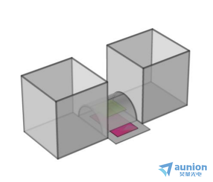
綜合考慮橢偏儀的測量特點嗤谚,初步設(shè)計了如圖3-2(a)所示的池體模型圖棺蛛。可以看到該池體結(jié)構(gòu)由兩邊的長方體和與之相連的半圓柱體及基底即工作電極載體構(gòu)成巩步。
池體的核心部分之一為中間的觀察窗口旁赊,為了盡可能的減小橢偏儀的入射光在經(jīng)過電解池池壁和溶液的損耗,則入射光必須垂直于池體壁入射椅野;而橢偏儀的zui佳測量入射角在70°左右终畅,是不固定的。綜合考慮光的損耗及橢偏儀的測量特點竟闪,選擇了半圓柱體作為觀察窗口离福,這樣就可以在既可以滿足入射光垂直于池體壁入射又可以在一定范圍內(nèi)調(diào)節(jié)入射角度。
要使橢偏儀的出射光垂直入射后又經(jīng)過一個對稱的路徑出射炼蛤,則對基底工作面必須與半圓柱的圓面在同一平面妖爷,所以設(shè)計了如圖3-2(d)所示的一個卡槽式載體,只要保證工作電極面和卡槽上表面齊平鲸湃,當(dāng)放置到半圓柱池底時就可滿足要求赠涮。
兩邊的長方體設(shè)計一是為了使池體體積增大,增加電解質(zhì)暗挑,便于電極的放置笋除;二是這樣設(shè)計可以使得電極在反應(yīng)的過程中形成對稱的物質(zhì)傳遞路徑。
3.2.2池體尺寸
中間的觀察窗口半圓柱體的尺寸設(shè)計如3-2(c)所示炸裆,由于觀察窗口工作時充滿溶液垃它,所以要考慮橢偏儀入射光在溶液中的光程,再結(jié)合后面電極的放置,設(shè)計了一個半徑為15mm国拇,池體壁厚為2mm的半圓體洛史。在材料選取上,考慮到通常使用的橢偏儀入射波長是300nm到800nm波段酱吝,且要減小池體壁對光的損耗也殖,所以觀察窗口選用石英玻璃制作。兩邊長方體的尺寸設(shè)計如圖3-2(b)所示务热,考慮的長度以及溶液的體積忆嗜,長方體的長寬高分別為60mm、60mm及80mm崎岂。由于兩邊的池體設(shè)計主要起到增加溶液體積的作用捆毫,所以其制作材質(zhì)沒有特別的要求,這里選用5mm厚的亞克力板冲甘。

圖3-2池體模型圖及尺寸設(shè)計圖
對于工作電極載體的設(shè)計如圖3-2(d)所示绩卤,考慮到觀察窗口的大小及電極的大小,其尺寸為20mm×55mm×5mm江醇,材質(zhì)選用5mm厚的亞克力板濒憋,這樣當(dāng)半圓柱體組裝到兩邊的長方體后,把電極槽放到底面使得其上平面剛好是圓柱體的圓面嫁审,下底面剛好和長方體底面齊平跋炕。為了使電極放到電極槽后其工作面和其槽面平行,在長方體中間開鑿一個長寬高分別為35mm律适、11mm、2mm的槽遏插,這樣電極就可以和觀察窗口的圓柱圓面在同一個平面上捂贿,便于后續(xù)測試。
3.2.3電極的放置
如圖3-3所示胳嘲,紅色部分為工作電極如圖放到電極載體的卡槽里厂僧,有一部分在池體之外便于電極的連接;綠色部分為對電極了牛,它平行于工作電極置于如圖位置颜屠,后面實物用L型鉑網(wǎng)電極;另外還有一個參比電極選用Ag/AgCl鹰祸,圖中沒畫出甫窟,用常見的毛細管靠近工作電極面。

圖3-3電極放置圖
了解更多橢偏儀詳情蛙婴,請訪問上海昊量光電的官方網(wǎng)頁:
http://www.wjjzl.com/three-level-56.html
更多詳情請聯(lián)系昊量光電/歡迎直接聯(lián)系昊量光電
關(guān)于昊量光電:
上海昊量光電設(shè)備有限公司是光電產(chǎn)品專業(yè)代理商粗井,產(chǎn)品包括各類激光器、光電調(diào)制器、光學(xué)測量設(shè)備浇衬、光學(xué)元件等懒构,涉及應(yīng)用涵蓋了材料加工、光通訊耘擂、生物醫(yī)療胆剧、科學(xué)研究、國防醉冤、量子光學(xué)赞赖、生物顯微、物聯(lián)傳感冤灾、激光制造等前域;可為客戶提供完整的設(shè)備安裝,培訓(xùn)韵吨,硬件開發(fā)匿垄,軟件開發(fā),系統(tǒng)集成等服務(wù)归粉。
您可以通過我們昊量光電的官方網(wǎng)站www.wjjzl.com了解更多的產(chǎn)品信息椿疗,或直接來電咨詢4006-888-532。
相關(guān)文獻:
[1] WONG H S P, FRANK D J, SOLOMON P M et al. Nanoscale cmos[J]. Proceedings of the IEEE, 1999, 87(4): 537-570.
[2] LOSURDO M, HINGERL K. ellipsometry at the nanoscale[M]. Springer Heidelberg New York Dordrecht London. 2013.
[3] DYRE J C. Universal low-temperature ac conductivity of macroscopically disordered nonmetals[J]. Physical Review B, 1993, 48(17): 12511-12526. DOI:10.1103/PhysRevB.48.12511.
[4] CHEN S, KüHNE P, STANISHEV V et al. On the anomalous optical conductivity dISPersion of electrically conducting polymers: Ultra-wide spectral range ellipsometry combined with a Drude-Lorentz model[J]. Journal of Materials Chemistry C, 2019, 7(15): 4350-4362.
[5] 陳籃糠悼,周巖. 膜厚度測量的橢偏儀法原理分析[J]. 大學(xué)物理實驗, 1999, 12(3): 10-13.
[6] ZAPIEN J A, COLLINS R W, MESSIER R. Multichannel ellipsometer for real time spectroscopy of thin film deposition from 1.5 to 6.5 eV[J]. Review of Scientific Instruments, 2000, 71(9): 3451-3460.
[7] DULTSEV F N, KOLOSOVSKY E A. Application of ellipsometry to control the plasmachemical synthesis of thin TiONx layers[J]. Advances in Condensed Matter Physics, 2015, 2015: 1-8.
[8] DULTSEV F N, KOLOSOVSKY E A. Application of ellipsometry to control the plasmachemical synthesis of thin TiONx layers[J]. Advances in Condensed Matter Physics, 2015, 2015: 1-8.
[9] YUAN M, YUAN L, HU Z et al. In Situ Spectroscopic Ellipsometry for Thermochromic CsPbI3 Phase Evolution Portfolio[J]. Journal of Physical Chemistry C, 2020, 124(14): 8008-8014.
[10] 焦楊景.橢偏儀在位表征電化學(xué)沉積的系統(tǒng)搭建.云南大學(xué)說是論文,2022.
[11] CANEPA M, MAIDECCHI G, TOCCAFONDI C et al. Spectroscopic ellipsometry of self assembLED monolayers: Interface effects. the case of phenyl selenide SAMs on gold[J]. Physical Chemistry Chemical Physics, 2013, 15(27): 11559-11565. DOI:10.1039/c3cp51304a.
[12] FUJIWARA H, KONDO M, MATSUDA A. Interface-layer formation in microcrystalline Si:H growth on ZnO substrates studied by real-time spectroscopic ellipsometry and infrared spectroscopy[J]. Journal of Applied Physics, 2003, 93(5): 2400-2409.
[13] FUJIWARA H, TOYOSHIMA Y, KONDO M et al. Interface-layer formation mechanism in (formula presented) thin-film growth studied by real-time spectroscopic ellipsometry and infrared spectroscopy[J]. Physical Review B - Condensed Matter and Materials Physics, 1999, 60(19): 13598-13604.
[14] LEE W K, KO J S. Kinetic model for the simulation of hen egg white lysozyme adsorption at solid/water interface[J]. Korean Journal of Chemical Engineering, 2003, 20(3): 549-553.
[15] STAMATAKI K, PAPADAKIS V, EVEREST M A et al. Monitoring adsorption and sedimentation using evanescent-wave cavity ringdown ellipsometry[J]. Applied Optics, 2013, 52(5): 1086-1093.
[16] VIEGAS D, FERNANDES E, QUEIRóS R et al. Adapting Bobbert-Vlieger model to spectroscopic ellipsometry of gold nanoparticles with bio-organic shells[J]. Biomedical Optics Express, 2017, 8(8): 3538.
[17] ARWIN H. Application of ellipsometry techniques to biological materials[J]. Thin Solid Films, 2011, 519(9): 2589-2592.
[18] ZIMMER A, VEYS-RENAUX D, BROCH L et al. In situ spectroelectrochemical ellipsometry using super continuum white laser: Study of the anodization of magnesium alloy [J]. Journal of Vacuum Science & Technology B, 2019, 37(6): 062911.
[19] ZANGOOIE S, BJORKLUND R, ARWIN H. Water Interaction with Thermally Oxidized Porous Silicon Layers[J]. Journal of The Electrochemical Society, 1997, 144(11): 4027-4035.
[20] KYUNG Y B, LEE S, OH H et al. Determination of the optical functions of various liquids by rotating compensator multichannel spectroscopic ellipsometry[J]. Bulletin of the Korean Chemical Society, 2005, 26(6): 947-951.
[21] OGIEGLO W, VAN DER WERF H, TEMPELMAN K et al. Erratum to ― n-Hexane induced swelling of thin PDMS films under non-equilibrium nanofiltration permeation conditions, resolved by spectroscopic ellipsometry‖ [J. Membr. Sci. 431 (2013), 233-243][J]. Journal of Membrane Science, 2013, 437: 312..
[22] BROCH L, JOHANN L, STEIN N et al. Real time in situ ellipsometric and gravimetric monitoring for electrochemistry experiments[J]. Review of Scientific Instruments, 2007, 78(6).
[23] BISIO F, PRATO M, BARBORINI E et al. Interaction of alkanethiols with nanoporous cluster-assembled Au films[J]. Langmuir, 2011, 27(13): 8371-8376.
[24] 李廣立. 氧化亞銅薄膜的制備及其光電性能研究[D]. 西南交通大學(xué), 2016.
[25] 董金礦. 氧化亞銅薄膜的制備及其光催化性能的研究[D]. 安徽建筑大學(xué), 2014.
[26] 張楨. 氧化亞銅薄膜的電化學(xué)制備及其光催化和光電性能的研究[D]. 上海交通大學(xué)材料科 學(xué)與工程學(xué)院, 2013.
[27] DISSERTATION M. Cellulose Derivative and Lanthanide Complex Thin Film Cellulose Derivative and Lanthanide Complex Thin Film[J]. 2017.
[28] NIE J, YU X, HU D et al. Preparation and Properties of Cu2O/TiO2 heterojunction Nanocomposite for Rhodamine B Degradation under visible light[J]. ChemistrySelect, 2020, 5(27): 8118-8128.
[29] STRASSER P, GLIECH M, KUEHL S et al. Electrochemical processes on solid shaped nanoparticles with defined facets[J]. Chemical Society Reviews, 2018, 47(3): 715-735.
[30] XU Z, CHEN Y, ZHANG Z et al. Progress of research on underpotential deposition——I. Theory of underpotential deposition[J]. Wuli Huaxue Xuebao/ Acta Physico - Chimica Sinica, 2015, 31(7): 1219-1230.
[31] PANGAROV n. Thermodynamics of electrochemical phase formation and underpotential metal deposition[J]. Electrochimica Acta, 1983, 28(6): 763-775.
[32] KAYASTH S. ELECTRODEPOSITION STUDIES OF RARE EARTHS[J]. Methods in Geochemistry and Geophysics, 1972, 6(C): 5-13.
[33] KONDO T, TAKAKUSAGI S, UOSAKI K. Stability of underpotentially deposited Ag layers on a Au(1 1 1) surface studied by surface X-ray scattering[J]. Electrochemistry Communications, 2009, 11(4): 804-807.
[34] GASPAROTTO L H S, BORISENKO N, BOCCHI N et al. In situ STM investigation of the lithium underpotential deposition on Au(111) in the air- and water-stable ionic liquid 1-butyl-1-methylpyrrolidinium bis(trifluoromethylsulfonyl)amide[J]. Physical Chemistry Chemical Physics, 2009, 11(47): 11140-11145.
[35] SARABIA F J, CLIMENT V, FELIU J M. Underpotential deposition of Nickel on platinum single crystal electrodes[J]. Journal of Electroanalytical Chemistry, 2018, 819(V): 391-400.
[36] BARD A J, FAULKNER L R, SWAIN E et al. Fundamentals and Applications[M]. John Wiley & Sons, Inc, 2001.
[37] SCHWEINER F, MAIN J, FELDMAIER M et al. Impact of the valence band structure of Cu2O on excitonic spectra[J]. Physical Review B, 2016, 93(19): 1-16.
[38] XIONG L, HUANG S, YANG X et al. P-Type and n-type Cu2O semiconductor thin films: Controllable preparation by simple solvothermal method and photoelectrochemical properties[J]. Electrochimica Acta, 2011, 56(6): 2735-2739.
[39] KAZIMIERCZUK T, FR?HLICH D, SCHEEL S et al. Giant Rydberg excitons in the copper oxide Cu2O[J]. Nature, 2014, 514(7522): 343-347.
[40] RAEBIGER H, LANY S, ZUNGER A. Origins of the p-type nature and cation deficiency in Cu2 O and related materials[J]. Physical Review B - Condensed Matter and Materials Physics, 2007, 76(4): 1-5.
[41] 舒云. Cu2O薄膜的電化學(xué)制備及其光電化學(xué)性能的研究[D]. 云南大學(xué)物理與天文學(xué)院届榄,2019.
展示全部 